Lesion in fatty liver
Kouseiren Takaoka Hospital
Dr. Yasushi Horichi, Dept. of Radiology
DATE : 2021
Introduction

Patient’s background
Male; 50s; body weight: 79.4 kg; lesion in fatty liver
Assessment objectives
August 12, 2020: Right high orchidectomy (T1bN0M0) performed for seminoma.
April 27, 2021: Follow-up computed tomography (CT) showed a new, faint, high-absorption region 1 cm in diameter in the periphery of S7 of the fatty liver.
Hepatic metastasis was suspected, so EOB-MRI was performed on May 17, 2021.
Contrast agent used
Gadoxetate disodium(Gd-EOB-DTPA) injection, 0.1 mL/kg
Case explanation
After surgery for seminoma of the right testis, the patient was monitored with simple CT every 4 months.
The original condition was severe fatty liver, but on April 27, 2021, a lesion that had not previously been found by CT was found. It was a faint, high-absorption region, approximately 1 cm in diameter, in the periphery of S7 of the liver, and was suspected of being a metastatic hepatic tumor.
EOB-MRI performed as a thorough examination gave negative results for metastatic and primary hepatic tumors, and enabled the diagnosis of focal spared area.
Follow-up CT performed on August 23, 2021, showed the high-absorption region to have disappeared.
Imaging findings

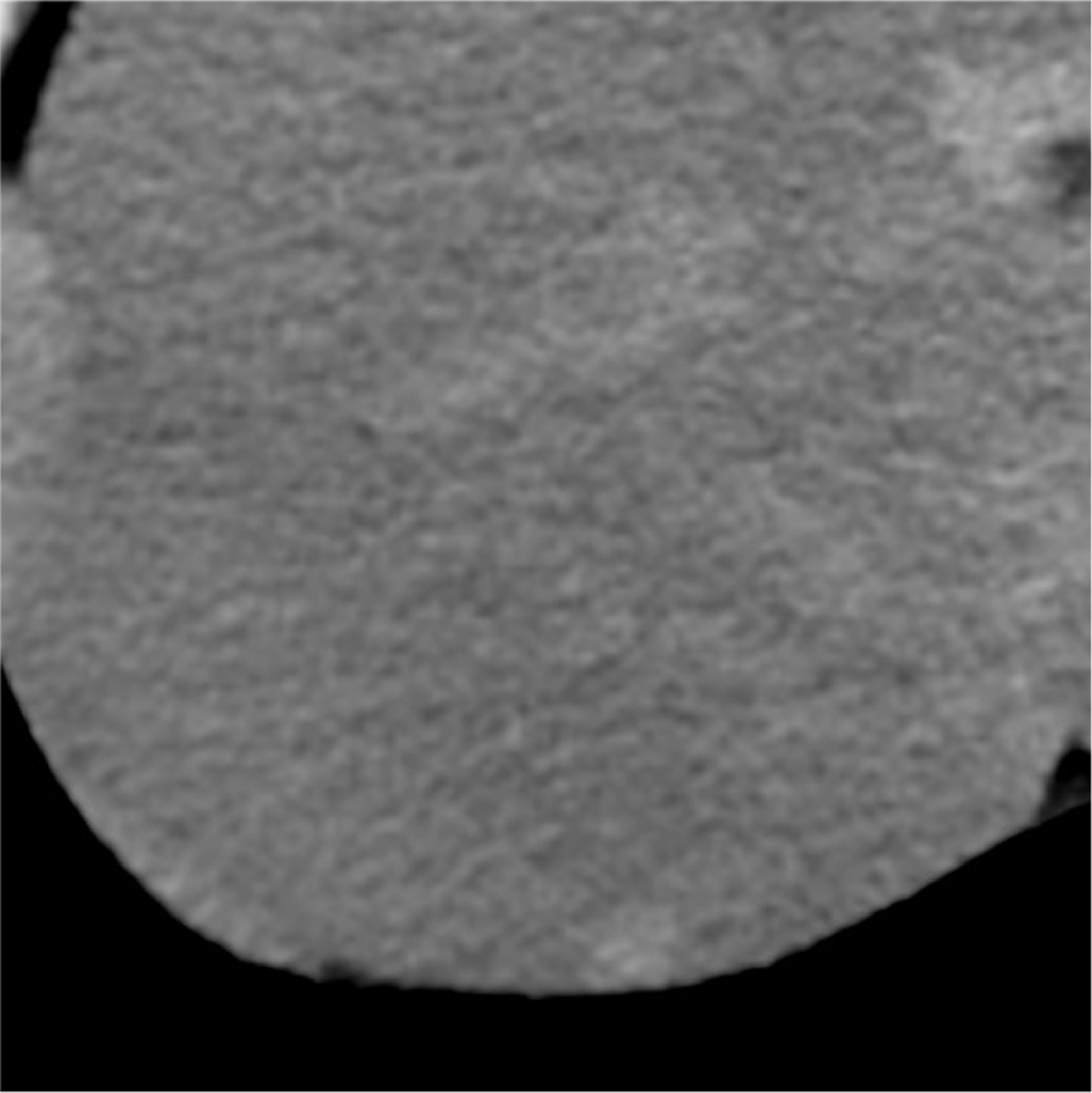
Simple CT showed fatty liver, and a faint high-absorption region approximately 1 cm in diameter in the periphery of S7.
Fig. 1. Simple CT

T1-, T2- and diffusion-weighted imaging showed isosignals, and no abnormalities were found.
Fig. 2. T1-, T2- and diffusion-weighted imaging

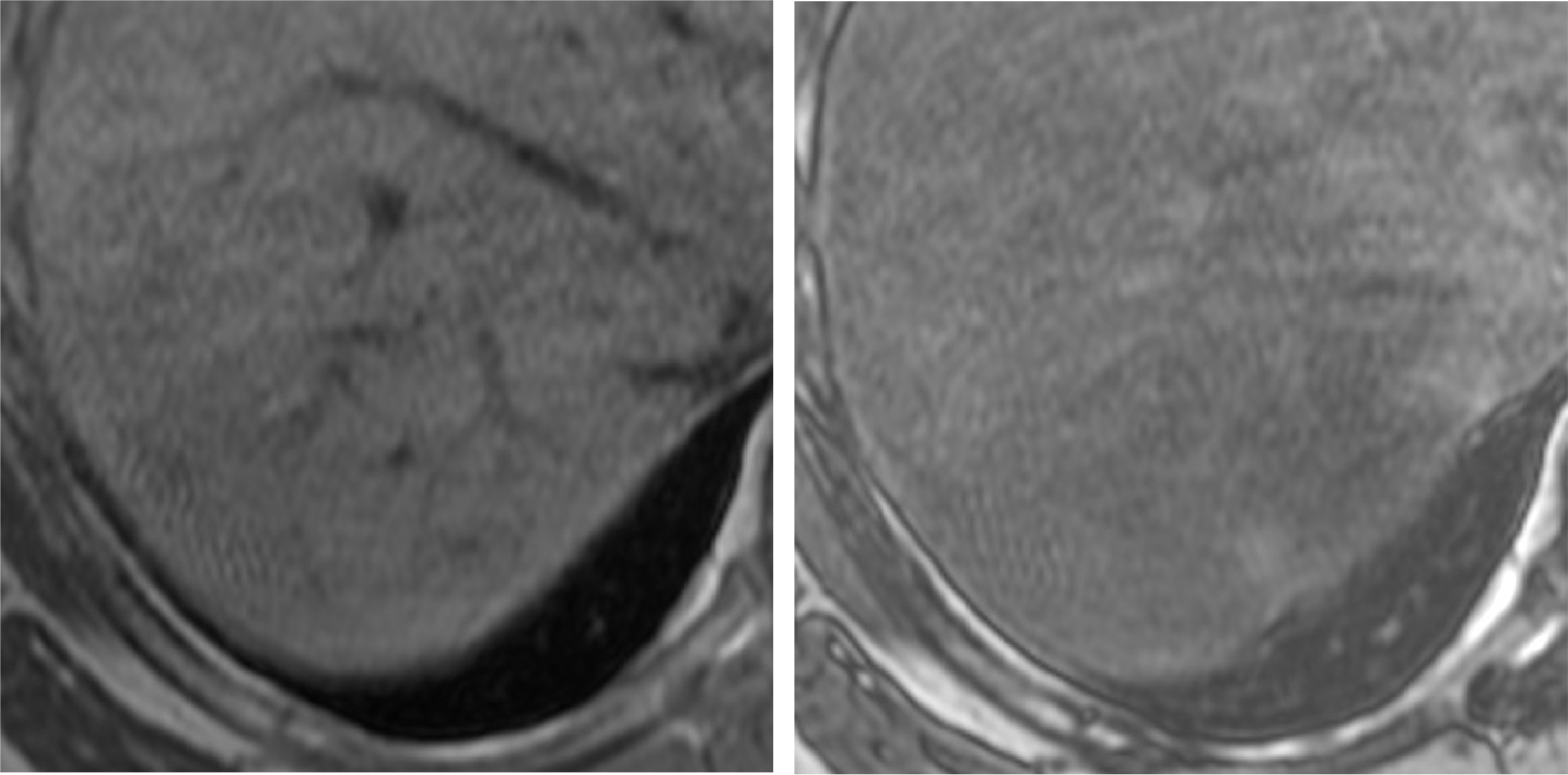
Whereas an isosignal was shown with in-phase imaging, with opposed-phase imaging the lesion region showed a faint high signal associated with a decreased signal in the surrounding fatty liver.
Fig. 3. T1-weighted imaging (in phase and opposed phase)

Faint dark staining was found from the arterial-dominant phase to the portal-dominant phase of dynamic imaging.
Fig. 4. Dynamic imaging

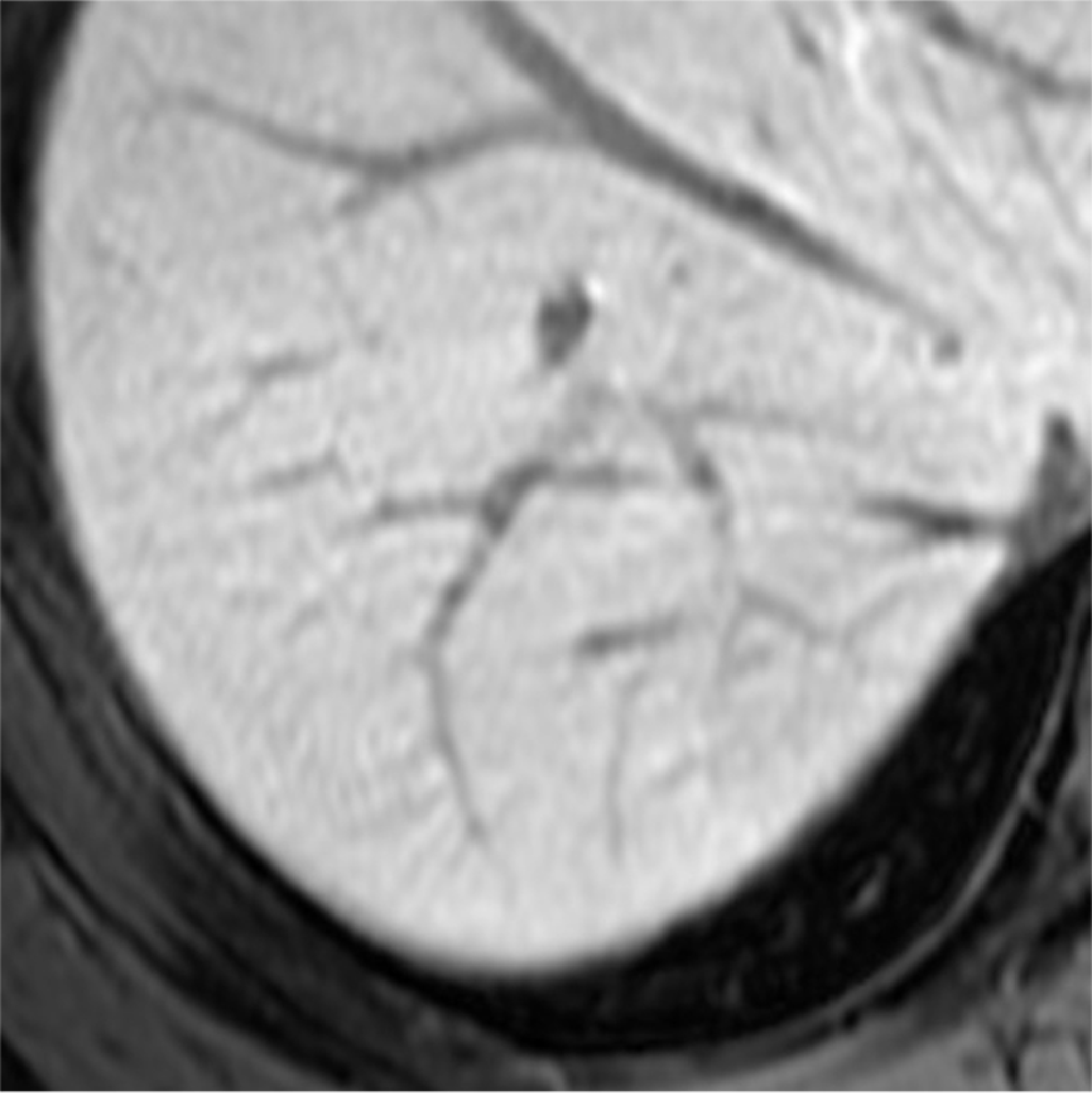
Even in the hepatobiliary phase, an isosignal was shown.
Fig. 5. Hepatobiliary phase
Photography protocol
| Imaging type | Photography sequence | TE (msec) | TR (msec) | TI (msec) | FA (deg) | Fat sat (type) | ETL (number) | Holding breath (yes/no) |
| Dual echo | ー | ー | ー | ー | ー | ー | ー | ー |
| Contrast agent administration | ||||||||
| Dynamic | Dynamic SmartPrep | 1.5 | 3.1 | 5.0 | 12.0 | DIXON | ー | Yes |
| DWI | DWI Resp b1000 | 53 | 13333.3 | ー | 90 | ー | ー | No |
| T2WI | T2 FS Resp PROPELLER | 78.9 | 17142.9 | ー | 125 | chemi sat | 26 | No |
| HBP | LAVA-FLEX | 1.7 | 3.8 | ー | 12.0 | DIXON | ー | Yes |
| in phase | LAVA-FLEX | 2.2 | 3.8 | ー | 12.0 | ー | ー | Yes |
| out of phase | LAVA-FLEX | 1.1 | 3.8 | ー | 12.0 | ー | ー | Yes |
| Imaging type | NEX (calculation number) | Slice thickness (mm) | FOV (mm) | Phase direction (step number) | Read direction (matrix number) | Slice Gap (mm) | Slice number |
| Dual echo | ー | ー | ー | ー | ー | ー | ー |
| Contrast agent administration | |||||||
| Dynamic | 0.7 | 4 | 360 | 512 | 512 | 0 | 100 |
| DWI | 2 | 5 | 360 | 256 | 256 | 1 | 35 |
| T2WI | 1.5 | 5 | 360 | 512 | 512 | 1 | 35 |
| HBP | 0.7 | ー | 360 | 512 | 512 | 0 | 100 |
| in phase | 0.7 | 4 | 360 | 512 | 512 | 0 | 100 |
| out of phase | 0.7 | 4 | 360 | 512 | 512 | 0 | 100 |
Devices used and contrast conditions
| MRI device | GE DISCOVERY MR750 |
| Automatic injection device | Sonic Shot GX (Nemoto Kyorindo) |
| Workstation | GE Advantage Workstation |
| Contrast conditions | Dose (mL) | Administration rate (mL/s) | Photography timing | |
| Gadoxetate disodium(Gd-EOB-DTPA) | 7.8 | 1 | smart prep. | |
| Physiological saline solution for flushing | 10 | 1 |
Usefulness of Gadoxetate disodium(Gd-EOB-DTPA) contrast MRI with this patient
With fatty liver, a focal spared area (i.e. a region with localized absence of fat deposition) associated with non-portal venous circulation is often found in loci such as surrounding the gallbladder bed, the portal region, and/or the region adjacent to the ligamentum teres hepatis in the periphery of the medial segment.
Single or multiple nodular spared areas are also sometimes found other than in the above typical loci, and are difficult to differentiate from primary and metastatic hepatic tumors by CT and ultrasonography.
Although MRI involving T2-weighted imaging, T1-weighted imaging, chemical-shift imaging, and diffusion-weighted imaging can be used for approximate assessment, EOB, a liver-specific contrast agent, is very useful because it facilitates visual assessment of the presence or absence of hepatocytes.
With the present patient, a faint dark-stained region consistent with the lesion was found in the early phase of dynamic imaging, but localized blood-flow abnormalities such as intrahepatic arterio-portal shunt may be suggested to be a factor in formation of the focal spared area.
- *The case introduced is just one clinical case, so the results are not the same as for all cases.
- *Please refer to the Package Insert for the effects and indications, dosage and administration method, and warnings, contraindications, and other precautions with use.


